![]()
![]()
![]()
![]()
A key task of the Research Department of Neuroscience is to further strengthen the existing excellent research in neuroscience and cognitive science and to provide state-of-the-art equipment. In recent years, numerous devices have been acquired for interdisciplinary research projects. The equipment acquired by the RDN is generally available to all members of the department, as well as to external interested researchers on request, if there is free capacity.
In addition to the infrastructure belonging directly to the RDN, members of the RDN also have access to other equipment within their respective faculties and their own working groups. If these devices can also be used by other groups, they are listed with the respective information.
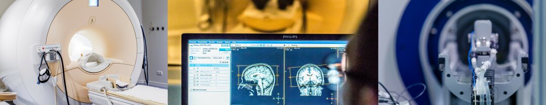
The MRI-scanner (Biospec 70/30 USR, Bruker, funded by Stiftung Mercator) is especially configured for MRI-studies of small mammals such as rodents and pigeons. Detailed pictures of brain structures in high contrast can be obtained based on the 7 tesla magnetic field strength. This high resolution is for example very important for experiments that aim at covering physiological to cognitive topics.
Contact:
Prof. Dr. Onur Güntürkün
Prof. Dr. Denise Manahan-Vaughan
Responsible for devices:
Dr. Xavier Helluy, MR physicist
Location:
Scanner Center RUB, building NI/NT
The 3 Tesla magnetic resonance imaging scanner (MAGNETOM Prisma, NX System, Siemens) was purchased in 2022 as part of a large-scale equipment proposal from the DFG. The MRI is designed in particular for neuroscientific questions using hardware and software equipment for optimized imaging of the human head and cervical, thoracic and lumbar spine, diffusion-weighted imaging, susceptibility weighted imaging, DTI examinations, BOLD-fMRI examinations and arterial spin labeling as well as spectroscopy.
The MRI scanner is located in a separate area on the first floor of the Interdisciplinary Institute for Teaching and Research (IFL) at the Ruhr University Bochum, which is located on the premises of the Katholisches Klinikum Bochum and is operated by the latter. In addition to the room for the scanner, the scanner area comprises a technical room, a control area (console and monitoring room) including two changing rooms and two examination/preparation rooms.
The use of the scanner is governed by user regulations. A steering committee regulates all matters relating to the use of the MRI scanner for research purposes.
Equipment includes:
Contact and responsible for devices:
Dr. Marco Meixner
Prof. Dr. Carsten Lukas
Dr. Barbara Bellenberg
Location:
Interdisziplinäres Institut für Forschung und Lehre (IFL) of Ruhr University Bochum at the St. Josef-Hospital Bochum, Gudrunstraße 56, 44791 Bochum, first floor
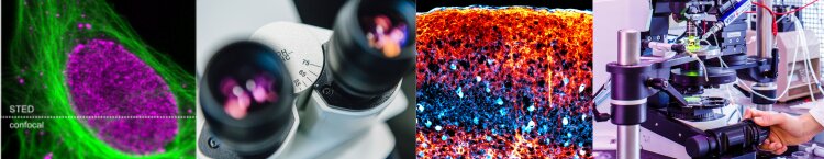
The STED microscope is based on the setup published by Bückers et al. (Optics Express 2011, 19(4), p.3130. DOI: 10.1364/OE.19.00313032011).
The light source of this setup is a Fianium ALP super-continuum laser offering a super-continuum as well as two beams for STED at 711nm and 745nm. The STED microscope is integrated into a commercial, automated epifluorescent microscope (iMIC, Till Photonics, today FEI Munich) which allows taking overview images. The filter sets currently available for the epifluorescent microscope allow recording the following dyes:
Objectives for the epifluorescent overview mode are Olympus 4× and 20× long working distance objectives. The epifluorescent microscope is built in a heating chamber that allows temperature control and controlling the CO2-level of the atmosphere, allowing for live cell recordings. STED imaging is possible with one orange-red and a second far-red dye.
Objective for STED recording is an Olympus APON60XOTIRF and confocal pinholes are 75µm in diameter, however, pinholes can be changed. The microscope is equipped with a Yanus IV beam scanner (Till Photonics, today FEI munich).
Contact and responsible for devices:
NN, please contact rd-neuroscience@rub.de
Location:
RUBION, RUB, Building NI 06/135
The system is based on a Sutter moveable objective microscope (MOM) and equipped with a high-power Mai Tai laser system (Newport Spectra-Physics). The laser is tuneable to achieve excitation wavelength from 690 nm – 1020 nm. The system is designed for in-vitro as well as for in-vivo investigations and can be widely adapted to the user’s requirements.
Contact:
Prof. Dr. Denise Manahan-Vaughan
Location:
Neurophysiology, Medical Faculty, MA 4, RUB
The system (Leica TCS SP5) based on a a multiphoton laser and fix-stage microscope. The system is designed for following investigations and can be widely adapted to the user’s requirements:
Contact:
Prof. Dr. Denise Manahan-Vaughan
Location:
Neurophysiology, Medical Faculty, MA 4, RUB
In the Imaging Center of the Department of General Zoology and Neurobiology, a spectrometer and various microscopes are available for use by internal and external scientists.
Further information with a list of all available devices as well as the respective contact persons can be found directly on the Imaging Center page.
To address its research interests experimentally, the Winklhofer lab is using state-of-the-art techniques in cell biology, molecular biology, protein biochemistry, and advanced light microscopy, including super-resolution microscopy and live cell imaging.
Further information with a list of all available devices can be found directly on the Winklhofer lab.
MIC acts as a central and competent center for the planning and implementation of modern light and electron microscopic analyses of various projects and issues in the field of life sciences and is available to all RUB faculties. The department for light microscopy is chaired by Prof. Dr. Konstanze Winklhofer, EMMACF (Electron Mircoscopy Medical Analysis – Core Facility) by Prof. Dr. Ralf Erdmann and Prof. Dr. Carsten Theiß.
The core of the EEG-lab is the 128-channel-EEG/EP-caps system with a dynamic impedance control for recordings of brain activity during diverse behavioral tasks (including a Brain Vision-Analyzer 2.0-software package).
Contact:
Prof. Dr. Nikolai Axmacher
Prof. Dr. Onur Güntürkün
Location:
RUB, Faculty of Psychology, Biopsychology, IB 6/96 + 150
Further information with a list of all available Open Software can be found directly on the Biopsychology homepage.
Further information with a list of all available Open Software can be found directly on the Neuroinformatics homepage.
Further information with a list of available shareware can be found directly on the homepage of the group "Neural Basis of Learning".
Photo collage "Magnetic resonance imaging": SFB 874/ Susanne Troll
Photo collage "Microscopy": SFB 874/ Susanne Troll, Ruxandra Barzan
Photo collage "Electroencephalography (EEG) laboratory": SFB 874/ Susanne Troll
Photo collage "Open software and hardware": pixabay.com
Copyright © RDN 2025
Last update: Apr 10, 2025


